Gene Editing of Human Embryos – Science Highlights November 2018

What is “Gene-Editing?”

Is gene editing human embryos a positive scientific breakthrough for human health?

Or misuse of a powerful research tool?

This “Highlights” will outline the basic biology behind gene editing, followed by a description of the process in general and in human embryos, specifically.

The Basics

The genetic information of humans, collectively termed the “human genome,” is contained within 22 chromosomes plus either 2 “X” chromosomes in girls, or 1 “X” chromosome and 1 “Y” chromosome in boys.

Human genome: All of the genetic information needed for the embryonic development and adult function of a human being.

Each chromosome is two long strings of four deoxyribonucleic acid (DNA) units (Adenosine, Cytosine, Guanosine, Thymidine; A, C, G, T) attached to each other in a sequence specific for that gene. The two long strings are held together by attractions between the units, i.e., A in one string is attracted to T in the opposite string.

Chromosome: a long string of genes attached end to end and then folded with proteins in a specific way.

Each species has its own number of chromosomes, e.g. the genome of the laboratory mouse is divided among 20 chromosomes, even though the total amount of DNA is the same in each mouse and human cell, approximately 5 picograms. An onion also has 20 chromosomes, but they are an order of magnitude larger than human or mouse, with approximately 50 picograms of DNA per cell. So the amount of genetic information does not correspond to the complexity of the organism.

Importantly, there are two copies of each chromosome present in all cells (total of 46), except for sperm, which have only one copy. This becomes an important fact for gene editing.

The specific sequence of A, C, G, T units is interpreted in two steps. First by assembling a copy of the gene sequence to serve as a template, and secondly by stringing amino acids together to generate the sequence specified by the template. The process is an engineering marvel that takes place billions of times every day in cells throughout the body. The gene’s code for a specific protein is the order of combinations of three of the four A, C, G, T units specific for each amino acid.

Gene: A specific sequence of A, C, G, T units that instruct the sequence of amino acids that comprise a specific protein. Humans have 20- to 25 thousand genes

Picture a train moving down a track. The two rails for the wheels are the two strands of DNA comprised of series of A, C, G, T units attached to each other and attracted to the opposite strand for stability. As the engine wheels pass by the units, the sequence for the emerging single stranded template is “read” in the car behind that contains a stockpile of A, C, G, U ribonucleic acid (RNA) units that are strung together in the same sequence as the gene. Behind the train car synthesizing the template is another car full of amino acids and the enzymes that string them together to create the protein specified by the gene.

The accuracy of the cellular machinery to “translate” gene sequences into the amino acid sequences for functioning protein molecules is both extraordinary and essential for normal cell functions.

For example, consider the gene that codes for a protein responsible for tissue rejection, beta 2 microglobulin, B2M. It is part of chromosome 15. The single stranded RNA template copy of the gene is 1675 units long and the protein it codes for contains 119 amino acids. The B2M protein begins with the string of eight amino acids linked together like beads: Methionine- serine- arginine- serine- valine- alanineleucine- alanine… which corresponds to gene sequence ATG TCT CGC TCC GTG GCC TTA GCT… This example also illustrates that each amino acid can have more than one triplet code, e.g. the triplet codes for serine are both TCT and TCC. This provides an important buffer for the specified amino acid should a T- to C- mutation occur in one of the codes for serine. And it also illustrates why everyone’s DNA sequences are not identical.

Moreover, to illustrate the importance of faithful replication of each chromosome every time a cell multiplies, if one unit were lost in the middle of the above sequence for B2M, e.g. a C, a “frame shift” would occur, and the sequence would become ATG TCT CGC TCG TGG CCT TAG… This sequence would code for methionine- serine- arginine- serinetryptophan- proline- followed by the “stop” triplet, TAG. Hence, no B2M protein would be synthesized as a result of a deletion of a single C.

Not only is translating the code into specific proteins essential to normal cell function, creating accurate and complete copies of all 46 chromosomes each and every time a cell multiplies is also essential to normal body functions.

Protein translation: The process of stringing together amino acids according to the sequence of A, C, G, T units in the gene

Because each amino acid is specified by a triplet sequence, if even one A or C or G or T is accidentally eliminated during replication of the chromosome, it would result in a “frame shift” in the triplet codes, as described in the B2M example.

This possibility is thought to be the reason for the second copy of each chromosome — as insurance that at least one copy of each gene will be available for the cell to use for essential processes. This is not the case for the X and Y chromosomes in males, which is why the disease hemophilia occurs in men. The genes that code for the proteins responsible for blood to clot following an injury are on the X chromosome. There is no back-up in men for mutations in X-chromosome genes, so such mutations result in loss of key blood clotting factors, hence hemophilia.

But there are extensive “gene repair” systems in every cell to correct mutations as they occur. Most mutations are probably due to the complexity of the enzyme systems themselves, others result from the relentless bombardment from gamma rays experienced by everything on earth. Every change in a gene sequence can be termed “gene editing,” whether or not it is repaired.

Naturally Occurring Gene Edits

No two individuals have exactly the same gene sequences because multiple sequences code for the same amino acid. This is the basis for DNA teststo prove paternity or predict ancestry. Most of the gene variations do not change the proteins they code for, but some do, such as genes for eye and hair color and height.

Gene edit: A modification of a specific sequence of A, C, G, T units that instruct the sequence of amino acids that comprise a specific protein. The edit may or may not alter the amino acid sequence and the protein.

Therefore, fertilization of an egg, pollination of a flower, introduce gene edits in the offspring because of variations in the gene sequences of the two cells uniting.

Still other gene edits occur because of “transposable elements,” first described in corn by Barbara McClintock (1), Nobel Laureate in 1983. Such “transposable elements” are common in all life forms, approximately 45% of the human genome is transposable elements and their location in individual genomes is highly variable.

The most well-studied gene edits in humans are those that cause cancer, such as the breast cancer gene, BRCA, on chromosome 13. It codes an important enzyme in DNA repair. A mutation that results in a “frame shift,” as described above, results in no BRCA protein expression. Hence, its function to repair spontaneously occurring DNA mutations is inhibited, resulting in cells containing mutated DNA that lack the controls that limit cell multiplication, leading to uncontrolled cell expansion, the definition of cancer.

A more recently studied naturally occurring gene edit is the 32 gene unit deletion in CCR5 on chromosome 3.

CCR5: A member of the C-C chemokine receptor family that codes for the docking protein for the HIV virus on the surface of HIV target cells.

The mutation results in loss of
CCR5 protein on the surface of HIV target cells, rendering them resistant to HIV attachment and infection. This mutation naturally occurs in approximately 1.5% of humans.

Gene Edits for Research

Early gene editing experiments were accomplished by mating individuals with different traits. Two well known examples are Mendel’s famous red peas crossed to white peas to yield pink peas, and Mr. Little’s Fancy Mice, popular in the early 1900’s, bred for coat color, formed the basis of the Jackson Laboratory’s inbred mice to study genetic diseases.

Nobel Laureate Mario Capecchi (link to his AES talk on the BRF site) systematically studied the function of mouse genes by mutating them into silence, so called “knock-out” mice.

Knock-out mouse: A form of gene editing that resulted in mutation of specific genes to silence them in order to discover their importance to normal functions, such as fetal development in the mouse.

This was accomplished by flooding cultures of mouse embryonic stem cells with strands of synthetic DNA that could replace the normal gene with an edited copy during DNA replication. The edited gene sequence was designed to not guide the synthesis of the normal protein. Such gene edited cells were combined with early mouse embryos, ultimately becoming part of the tissues of the mouse, including occasionally sperm and eggs. Males with gene edited sperm were mated to females with gene edited eggs to produce offspring containing two copies of the edited, non functioning genes. Although laborious and time consuming, this approach has yielded highly valuable information about the normal functions of thousands of genes.

In the past 20 years, other less time consuming methods of silencing genes, or increasing their expression, have been developed, all with the goal of understanding their function in health and disease.

In 2013, the most recent method for gene editing was popularized by scientists at Stanford and MIT.

Knock-out mouse: A form of gene editing that resulted in mutation of specific genes to silence them in order to discover their importance to normal functions, such as fetal development in the mouse.

It is an adaptation of a naturally occurring defense mechanism that bacteria have against the viruses that invade them. Termed CRISPR/Cas, it is a complex between a protein that can cut DNA strands and a synthetic single-stranded RNA with a sequence of A, C, G, U that matches the gene being targeted. The simplicity and specificity of the system have rapidly led to a wide variety of applications among scientists world-wide.

Such targeted DNA cuts can edit the gene sequences so they no longer code for a functioning protein, analogous to the natural CCR5 mutation, or opening the DNA strands can allow the incorporation of synthetic DNA sequences into the cut site. This raises the exciting possibility of being able to repair defective human genes.

Gene Edits for Treatment of Disease

Most scientists have applied the CRISPR/Cas system to specific tissues or to stem cells. For example, it is theoretically possible to repair the X-chromosome mutations in liver cells so normal blood clotting factors can be produced by the liver (2).

Bedford Research scientists are applying the technology to edit B2M gene sequences in unfertilized eggs which are subsequently activated for stem cell derivation (link to article on website).

But more recently other scientists have applied CRISPR/Cas technology to human embryos. Last year a Portland Oregon research team reported their
efforts to repair a mutation in the gene MYBPC3 known to be associated with acute heart failure in young men (3). The 30-member team created embryos with sperm from a man carrying the mutated gene in half of his sperm. (It is important to note that this experiment is not possible in Massachusetts because the stem cell bill (MGLc 111L) specifically prohibits the creation of embryos for research purposes only.)

At the time of fertilization of eggs with the mutant sperm, the Oregon scientists also injected the CRISPR/Cas agents designed to home to the gene
mutation and insert “normal” DNA sequences. They reported the repair was successful in some embryos, but not all. Other research teams in New York and Australia replied to the report with their own interpretations of the results and all groups agreed much more work is needed to understand how to reliably edit genes in early human embryos.

Gene Edits for Enhancement

Earlier this week, a Chinese scientist reported the birth of twin girls whose genomes had been modified to silence the CCR5 gene (4). The birth was reported to be one of a series of human embryo experiments designed to render the offspring resistant to infection by HIV and to prove the principal that gene editing was possible — and perhaps beneficial— in human embryos. The work was not reported in a scientific format, so few scientists have had the opportunity to review the data in detail.

Several ethical concerns with this report, if true, have been raised. The gene editing was not performed to correct a known, serious medical issue in the embryos. It was performed to enhance resistance to HIV. A highly controversial idea. But a more practical problem with the work is the
possibility of “off-target” gene edits. Much research has been devoted to discover, and eliminate, the random edits that may occur at other than the gene locations being specifically targeted by the CRISPR/ Cas reagents. It is these potentially deleterious unintended consequences that must be addressed in order to protect the offspring produced.

The Future of Gene Editing

Gene editing is common in nature, and forms a basis for evolution itself. Some edits are positive, such as the mutation that leads to resistance to HIV infection; other edits are negative such as the ones causing hemophilia and heart failure.

Humans have been gene editing plants, animals, and each other by cross-breeding for millennia. Technology companies like Monsanto have harnessed natural plant gene editing systems to modify plant genes such as rice to produce Vitamin A and corn to resist infestation by worms. Termed “genetically modified organisms” such plants have caused heated world-wide debate, despite the fact that cross-breeding plants modifies many more genes in one breeding cycle than the Monsanto technology.

Nonetheless, every new technology that can have effects in subsequent generations — of plants, animals, or microorganisms — needs to be carefully reviewed and implemented in stages that can be monitored for unintended, deleterious consequences.

We cannot put this genie back in the bottle, but with reasoned approaches, humans can optimize the benefits and mitigate the dangers posed by gene editing.

Ann A Kiessling, PhD
(1) Carnegie Institute, 1948;
(2) http://hemophilianewstoday.com/crispr-cas9-hemophilia
(3) Nature, 2017, 548:413; (4) https://www.technologyreview.com/s/612472/rogue-chinesecrispr-scientist-cited-us-report-as-his-green-light/

In Memoriam of Dr Paul Winig

Dr. Paul Winig, a long time collaborator and benefactor of The Foundation passed away on September 9th, 2018. Dr. Winig was a key collaborator and founder of the Foundation’s SPAR program (originally the Assisted Reproduction Foundation), as well as a participant in the Stem Cell Research programs that have been performed since the early 2000s at the Foundation. A dedicated Obstetrician, gynecologist and researcher, he had a passion for being “behind the scenes” for medical science.

A full obituary of his extraordinary life and accomplishments is posted on the Boston Globe’s obituary website.

We are grateful to have known him and worked with him. He will be missed.

Bedford Research Foundation 2017 Newsletter

Read about all of the progress and the research that has occurred at the Foundation over the course of the past year, and a retrospective on the past 21! Dr. Kiessling outlines her vision for the upcoming year as well. Thank you for your support.


Bedford Research Foundation is TWENTY ONE

Founded in 1996 to conduct research that cannot be funded by the National Institutes of Health, Bedford Research scientists have achieved ground-breaking milestones!

See our Timeline of Milestones!


 Bedford Research Foundation’s work cannot be federally funded because of the Dickey-Wicker Amendment to the budget of the National Institutes of Health, put in place in 1996 and renewed annually. BRF scientists need private donations for research to develop “universal” stem cells for Everybody.
Donate Today!

BRF Research News

Our goal for 2017 was to improve the efficiency of a new technology, “gene editing” by CRISPR, that can precisely edit genes in eggs activated to become stem cells. BRF scientists accomplished this goal in a mouse model by developing new methods that improve the efficiency of CRISPR gene editing in mouse eggs from 10% to approximately 75%, with the added success of deriving stem cells from more than 50% of the gene edited, activated eggs.

Two genes were simultaneously targeted for editing:

(1) Just as Type “O” blood can be given to almost everyone, a “universal” stem cell could be missing the gene, B2M, responsible for the proteins on stem cells that cause immune rejection following transplantation. Such a “universal” stem cell could be transplanted into many individuals without leading to immune rejection. This is an essential step to the derivation of “off-theshelf” stem cells for everybody.

The 2017 mouse egg stem cell experiments by BRF scientists derived mouse stem cells missing B2M. This paves the way to translate the research to the derivation of universal stem cells from human eggs. Like blood banks, universal stem cell banks would be available in hospitals for acute treatments, such as heart attack, stroke and spinal cord injury.

(2) CRISPR gene editing can also mimic the natural mutation in 1% of humans that renders individuals resistant to infection by HIV, the virus that causes AIDS. The recent success in mouse eggs to eliminate the HIV receptor, CCR5, paves the way to deriving a library of universal human stem cells also resistant to HIV infection.

IF those cells can be developed into bone marrow stem cells, and IF those bone marrow stem cells will function normally, they could be utilized as a powerful treatment, perhaps a cure, for HIV disease.

New Research Program a Success in Mouse Stem Cells

Dr. Joel Lawitts microinjects CRISPR/Cas “gene editing” enzymes into mouse eggs to neutralize two genes at once: (1) the gene that leads to tissue rejection, and (2) the gene that allows HIV infection of cells. These are the first steps in generating off-the-shelf stem cells for everybody that are also resistant to HIV infection.

“Dr. Kiessling and her staff have shown their determination to tackle some of the most difficult health problems of our time.”
– Representative Ken Gordon

Dr. Robert EyreProstate Disease Research Update

Along with continuing patient recruitment into the prostate cancer screening project, BRF Scientists have developed methods for including specimens submitted to the laboratory for other types of testing. The goal of the project is to develop semen screening tests that improve diagnosis and staging of prostate cancer as well as reflect overall male health.


Alex HauserNew Member Of The Bedford Research Team

Alex Hauser graduated from UMass Lowell with a B.S. in Biology in 2017 and joined the BRF staff full time. Alex is focused on the SPAR program and also has an interest in gene editing technology and analysis of cell lines.


Dr. Bronte StoneOur New Science Advisor

Dr. Bronte Stone holds a PhD in Medicine and has directed the Laboratory of California Fertility Partners with over 30 years experience in human eggs and embryo development. Dr. Stone joins our scientific advisory board as we enter the human egg phase of our research.


Taylor MellenMeet Our Mass Life Science Center Intern

The Massachusetts Life Science Center sponsors internships in companies engaged in life science development in the Commonwealth. Taylor Mellen will graduate from UMass Lowell in 2018 with a B.S. Degree in Biology. We’ve enjoyed having Taylor with us working in the andrology lab services.


From the Director

The derivation of gene edited, universal, HIV-resistant human stem cells from unfertilized eggs will not be without controversy. Fortunately, we have meritorious individuals serving as our Ethics Advisory Board, our Human Subjects Committee and our Stem Cell Research Oversight Committee. Their guidance has helped us forge ahead into areas of stem cell development that larger institutions have shied away from because the work cannot be funded by our federal government. The “Dickey-Wicker Amendment” to the budget of the National Institutes of Health has been renewed annually and prohibits funds to be used for studies of unfertilized human eggs. We have for years believed unfertilized eggs  (“parthenotes”) will be a broadly applicable source of “universal” human stem cells for everybody. Since human egg research MUST be privately funded, progress depends entirely on private donations. BRF is uniquely positioned to push this exciting field forward, and we need everyone’s support!

Sincerely,

Ann A Kiessling, PhD
Director, Bedford Research Foundation

Who is Bedford Research Foundation?

Philanthropy Is The Key To Continued Progress

The average cost of each experiment is $90,000. Because much of our overhead is covered by fee-for-service laboratory tests, 92% of every dollar donated goes directly toward these experiments. This innovative funding model allows Bedford Research scientists greater flexibility to move quickly in promising new research directions.

Continued progress requires meeting our annual funding goal.

Donate Today!

Circadian Rhythms – Science Highlights October 2017

Circadian Rhythms in the Spotlight

The 2017 Nobel Prize in Physiology for circadian rhythm discoveries is exciting news for Bedford Research.

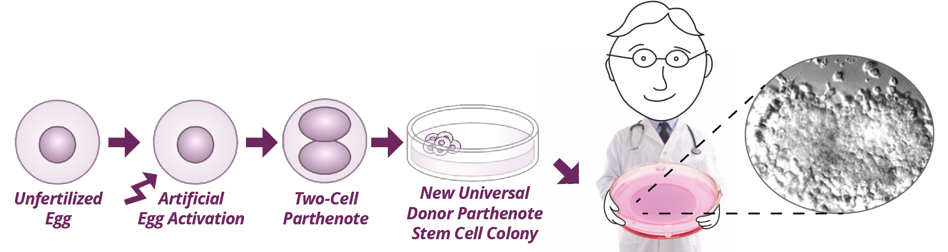
Almost a decade ago, Bedford Research scientists discovered circadian rhythm genes were turned on as early as three days (the 8-Cell stage) after a human egg was activated by sperm (1).

Although circadian rhythms were discovered over a century ago, their importance and their molecular basis is a fast-growing field in the past 20 years.  Bedford Research scientists became aware of circadian rhythm genes while researching ways to improve the efficiency of developing therapeutically valuable stem cells from human eggs activated without sperm (parthenotes).


Circadian Rhythm:  a behavior that repeats every 24 hours


The new Nobel Laureates, Jeffrey Hall and Michael Rosbash of Brandeis University, and Michael Young of Rockefeller University, simultaneously discovered a protein, “PER,” in fruit flies over 30 years ago that increased after dark and decreased during the day.  The key finding was that although the rise and fall of “PER” was entrained by cycles of light and dark, the circadian pattern persisted even after the fly was kept in solid darkness for several days.  Hence, production of the protein wasn’t directly stimulated by light exposure, but had become its own internal, circadian mechanism.

We now know that all cells are regulated by circadian rhythms, a fundamentally important process that controls both behavior, e.g. sleep, and cellular processes, such as release of hormones.

Mammalian circadian rhythm genes are different from those in fruit flies, and were discovered by several investigative teams in the 1990’s:

Takahashi and colleagues identified the mammalian gene, CLOCK, as essential for normal circadian rhythms. Mammalian cells have three “PER” genes:  PERIOD 1, -2, -3, plus two forms of a gene, CRYPTOCHROME discovered by Sancar and colleagues to form a complex with PERIOD.

Hogenesch, Ikea and Nomura discovered ARNTL (also known as BMAL), which was shown to form a complex with CLOCK by Weitz and colleagues, including Fred Davis, Bedford Research Advisor.

The picture has now emerged of an elegantly simple feed-back loop that takes 24 hours to complete, is thought to control about 12% of mammalian genes, and is maintained by stimuli from the supra-chiasmatic nucleus in the hypothalamus.


Core circadian oscillators:  CLOCK/ARNTL stimulates PER/CRY which feed-back inhibits CLOCK/ARNTL, which decreases PER/CRY allowing CLOCK/ARNTL to increase again, in a 24 hour cycle.


Bedford Research scientists, however, have more recently reported(2,3) that approximately 70% of key regulatory genes expressed at high levels at the 8-Cell stage of development are circadianly controlled(4), and that in contrast to the 8-cell stage, the core circadian oscillators in human stem cells in long term culture are silent, calling into question the normality of their responses during experiments.

There is a pressing need to support circadian rhythms during the derivation, long term culture and study of human stem cells.  Bedford Research scientists began to develop such methods a decade ago, the importance of which is supported by this year’s Nobel Prizes.  Hearty congratulations to Jeffrey Hall, Michael Rosbash and Michael Young.

(1) JARG 26:187; (2) JARG 27: 265; (3) SCD 25:160;    (4) circadb.hogeneschlab.org

Download the PDF

Job Opening: Scientific Director

The Bedford Research Foundation is seeking a scientific director to lead its program to derive parthenogenetic stem cells from unfertilized human eggs.  Experience with human eggs and stem cell derivation is required.  Proven success in obtaining grant funding and a strong publication record are also required.

Learn More

Bedford Research Foundation 2016 Newsletter

Read about all of the progress and the research that has occurred at the Foundation over the course of the past year, and a retrospective on the past 20! Dr. Kiessling outlines her vision for the upcoming year as well. Thank you for your support.


Bedford Research Foundation is TWENTY

Founded in 1996 to conduct research that cannot be funded by the National Institutes of Health, Bedford Research scientists have achieved ground-breaking milestones!

See our Timeline of Milestones!


 Bedford Research Foundation’s work cannot be federally funded because of the Dickey-Wicker Amendment to the budget of the National Institutes of Health, put in place in 1996 and renewed annually. BRF scientists need private donations for research to develop “universal” stem cells for Everybody.
Donate Today!

From the Director

It is finally all coming together…

My career in reproductive biology and AIDS began in 1983, with the goal of understanding the influence of viruses on early embryo development. Wonderful young scientists joined my laboratory for periods of training during the ensuing 33 years, and together we have made many discoveries that bring our laboratory skills to where they are today. We now have the foundation to begin to generate off-the-shelf stem cells for everybody.

Additionally, because we are a nimble institution, we were able to quickly change research direction to take advantage of a new technology, reported in 2013, that allows unprecedented precision in silencing genes. This advance has two immediately practical applications for our “off-the-shelf” stem cell research goals:

(1) It is now feasible to specifically silence the genes responsible for the proteins on cells that cause immune rejection. Just as Type “O” blood can be administered to almost everyone, such a neutralized cell could be transplanted into many individuals without leading to immune rejection. This would be a major step forward in generating “off-the-shelf” stem cells for everybody. Our successful experiments in mouse eggs pave the way to translate the work to stem cells from human eggs. Like blood banks, such a stem cell bank could be available in emergency rooms for acute treatments, such as heart attack, stroke and spinal cord injury.

(2) It is also feasible to replicate the natural mutation in 1% of humans that renders individuals resistant to infection by HIV, the virus that causes AIDS. The ability to precisely silence this gene without causing other changes in the cell, in the same way it is naturally inactivated in 1% of humans , paves the way to deriving a library of stem cells resistant to HIV infection. IF those cells can be developed into bone marrow stem cells, and IF those bone marrow stem cells will function normally, they could be utilized as a powerful treatment, perhaps a cure, for HIV disease.

To help guide the work, we have meritorious individuals serving as our Ethics Advisory Board, our Human Subjects Committee and our Stem Cell Research Oversight Committee. Their guidance has allowed us to forge ahead into areas of stem cell development that larger institutions have shied away from because the work cannot be funded by our federal government. The “Dickey-Wicker Amendment” to the budget of the National Institutes of Health is renewed annually and prohibits funds to be used for studies of unfertilized human eggs. We have for years believed unfertilized eggs (“parthenotes”) will be a broadly applicable source of human stem cells.

Human egg research MUST be privately funded, progress depends entirely on private donations. No federal dollars can be used to study activated human eggs or parthenote stem cells. BRF is uniquely positioned to push this exciting field forward! Thank you for your continued support.

Sincerely,

Ann A Kiessling, PhD
Director, Bedford Research Foundation

Who is Bedford Research Foundation?

Stem Cells for Every Body

Unfertilized eggs can be activated artificially (parthenogenesis) by an electrical jolt or chemical stimulation. Activated eggs (“parthenotes”) undergo early cell divisions similar to fertilized eggs, but cannot give rise to offspring. At the time of activation, a protein responsible for tissue matching can be silenced. “Universal donor” stem cells could then be isolated from such parthenotes. Similar to Type O blood, such “universal” stem cells could be available “off-the-shelf ” in emergency rooms for acute injuries, such as heart attack, stroke and spinal cord injury.

Philanthropy Is The Key To Continued Progress

The average cost of each experiment is $90,000. Because much of our overhead is covered by fee-for-service laboratory tests, 92% of every dollar donated goes directly toward these experiments. This innovative funding model allows Bedford Research scientists greater flexibility to move quickly in promising new research directions.

Continued progress requires meeting our annual funding goal.

Donate Today!

Volunteer of the Year

Deborah WeidmanWe are very grateful for the help from our Volunteer of the Year, Deborah Weidman. A Bedford High School student, she has been instrumental to our circadian rhythms in stem cells research this year. She is planning to use her experience with BRF in her career in biomedical engineering. Thank you Deborah!

Prostate Disease Research Update

Dr. Robert Eyre Patient recruitment into the prostate cancer screening project is ongoing, and Bedford Research Scientists have developed methods for including specimens submitted to the laboratory for other types of testing.  The goal of the project is to develop semen screening tests that improve diagnosis and staging of prostate cancer as well as reflect overall male health. Urologists from around the country have joined the research.

On twenty years of progress: Letter from our director

With the help of generous philanthropists and medical collaborators, Dr. Ann Kiessling founded the Bedford Research Foundation in 1996 to address a research need that could not be federally funded — how to help men infected by HIV through tainted blood transfusions have children without infecting their wives and babies. The first “Special Program for Assisted Reproduction,” or SPAR baby was born in 1998.  Ryan Schlosser, now 16 years old, visited the Bedford Research lab last summer. As of September, 2015, 246 babies SPAR babies have been born with all moms and babies testing negative for HIV.Ann Baby Ryan

In 1999, BRF responded to a new research need that could not be federally funded — the derivation of stem cells from unfertilized human eggs.  Bedford Research scientists spear-headed the world’s first ethics advisory board and medical team charged with the task of developing the “gold standard” for women volunteering to donate their eggs for research.

To be clinically feasible, at least 10% of activated, unfertilized human eggs must successfully develop into stem cells.  When initial experiments failed to reach this goal, BRF scientists collaborated with colleagues in Greece to discover what genes must be activate in unfertilized eggs to reach the 10% efficiency needed.  Our results, published in three landmark reports, have led to the information needed to resume the research.  As described in the cover story, the work will need eggs donated by women for research, a controversial topic being considered by BRF ethicists.

Human egg research must be privately funded.  Due to the National Institutes of Health budget no federal dollars can be used to study activated human eggs, or parthenote stem cells.  Bedford Research is uniquely positioned to push this field forward, and needs to add two additional scientists in 2016 to optimize progress. Progress depends entirely on private donations.

Check out more news in the BRF Fall 2015 Newsletter.

Save

Meet our new board members!

The Bedford Research Foundation welcomes two new board members to the Board of Trustees:  Larry LaFranchi, Ellen Sheehy and Scott Anderson.

Larry LaFranchi brings more than 30 years of business and entrepreneurial experience. His passion for health care and is a perfect match for the Foundation’s independent research goals, and his expertise in financial planning and consulting will benefit all aspects of research planning.

Ellen Sheehy is an experienced, analytical entrepreneur with broad experience in strategy development and implementation.  She has been a leader in the field of nonprofit healthcare for many years.

Scott Anderson is an experienced computer developer and technical science writer. He co-authored Human Embryonic Stem Cells with Dr. Kiessling.

Check out more news from the BRF Fall 2015 Newsletter.