A Faster Way to Study Stem Cells—With a Surprising Observation About Fertility

An improved method for deriving stem cells shortens the time to just a few weeks(rather than months)—changing how scientists can study stem cells, fertility, and mitochondrial health.

Developed at Bedford Research Foundation and presented in an award-winning research poster, this faster method for creating mouse embryonic stem cells has already revealed unexpected patterns in how cells with metabolic defects behave, opening the door to accelerated studies of age-related fertility decline.

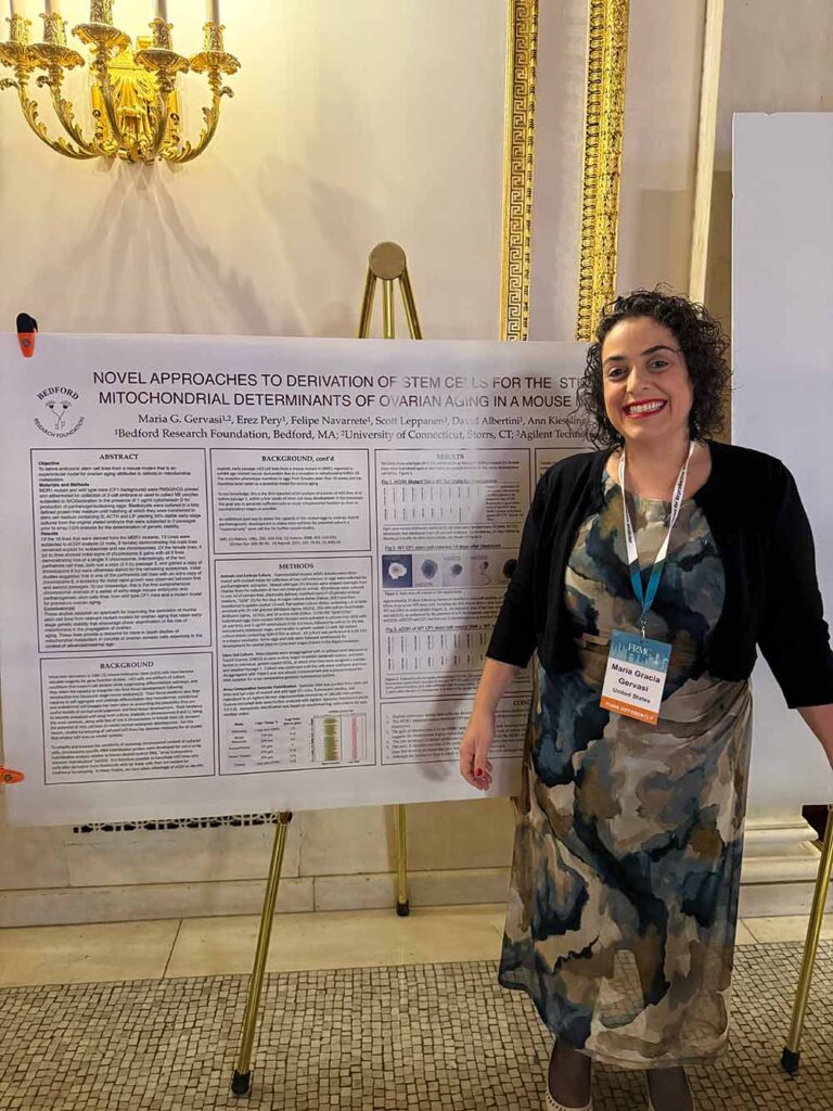
Dr. Maria G. Gervasi in front of the prize winning poster at The Foundation for Reproductive Medicine Conference 2025 in New York, Dec 5-7.

A Faster Protocol

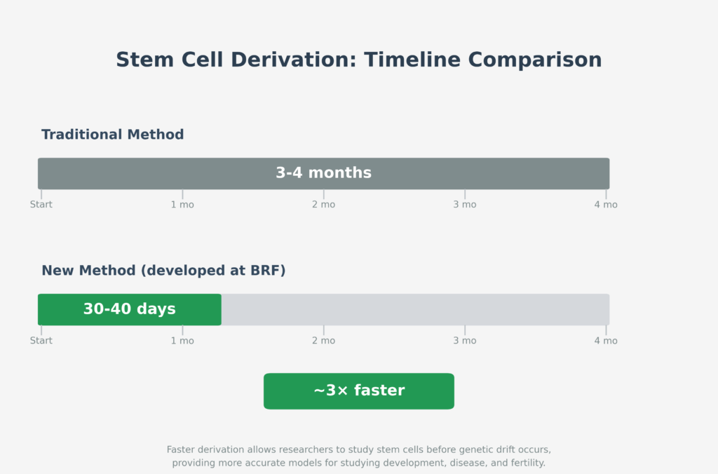
Creating stem cell lines from mouse embryos has been a foundational research technique since 1981—it helps us understand everything from genetic diseases to tissue development. But there’s been a catch: by the time you grow enough cells to study them (usually 3-4 months), they’ve changed genetically, potentially rendering them too different from the intended model to yield accurate insights.

BRF scientists developed a new technique that speeds this up dramatically—generating stable, analyzable stem cell lines in just 30-40 days. That means researchers can study cells while they’re still in a near-pristine state, before genetic drift sets in.

“We wanted to capture these cells as close to their natural embryonic state as possible,” says Dr. Ann Kiessling. “Fast enough that we’re seeing what’s really there, not what laboratory conditions have turned them into.”

Proving It Works: Stable Lines from Abnormal Embryos

The team put their technique to a rigorous test: embryos from mice with a specific metabolic defect that causes early ovarian aging in females. These weren’t normal embryos—they were known to be abnormal, representing challenging starting material for stem cell derivation.

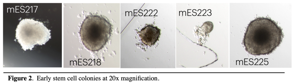
The faster protocol succeeded. Even with compromised embryos, the researchers generated stable, usable stem cell lines within the 30-40 day timeframe. This proves the technique is robust enough to handle difficult cases, not just ideal conditions.

The broader impact is significant: researchers across any field of stem cell biology now have a faster, more reliable method for establishing cell lines. And for those specifically studying mitochondrial metabolism and its effects on eggs and ovarian cells—particularly in the context of aging—these validated cell lines provide a ready-made model system that can be analyzed in weeks rather than months.

A Window Into Age-Related Fertility Decline

While validating their protocol, the researchers noticed something unusual in the genetic profiles of their stem cell lines.

The stem cells from female embryos lost an X chromosome much earlier and more frequently than previously documented. The cells from male embryos remained stable. Some female cell lines also gained extra copies of chromosome 8—and those cells grew faster, suggesting a possible compensation for their metabolic defects.

The patterns are intriguing—cells with metabolic defects showing specific types of genetic instability. If similar patterns occur in aging human eggs, understanding these changes could eventually point toward approaches for fertility preservation. For now, these findings raise important questions for future research.

This research was conducted by Maria G. Gervasi, PhD, Erez Pery, PhD, Felipe Navarrete, PhD, PhD, David Albertini, PhD, and Ann Kiessling, PhD at Bedford Research Foundation in collaboration with Scott Leppanen at Agilent Technologies and the University of Connecticut, and with support from the ESHE Fund.  

Breakthrough Study Demonstrates Viability of Ambient Temperature Transport for Human Oocytes, Unlocking New Research Opportunities

The study, titled “Ambient Temperature Transport of Human Oocytes: An Unexpected Research Resource,” explored the potential of immature oocytes from young egg donors at The World Egg and Sperm Bank in Phoenix, AZ. A total of 432 oocytes, collected from controlled ovarian stimulation cycles, were shipped overnight in a specialized transport medium designed to mimic the follicular environment. The medium included caffeine, dibutyryl cyclic-AMP, estrogen, progesterone, and, in some cases, zinc sulfate (ZnSO4). Remarkably, over 95% of the oocytes remained viable upon arrival, with many demonstrating meiotic progression or spontaneous activation after culture.

Key Findings:
High Viability: More than 95% of oocytes survived ambient temperature transport, whether supplemented with zinc or not, showcasing the robustness of the transport protocol.
Meiotic Competence: Transported oocytes exhibited meiotic progression, with some advancing to metaphase II or undergoing spontaneous activation, particularly when zinc was included in the transport medium.
Spontaneous Activation: Oocytes transported with zinc showed a higher incidence of spontaneous activation and cleavage activity, progressing to 2-, 4-, and even 8-cell stages during extended culture, as observed through time-lapse imaging.
Research Potential: The study highlights the potential of using “discard” oocytes—typically unsuitable for clinical use—as a valuable resource for studying oocyte biology and developing parthenogenetic stem cells, which could offer ethical and practical advantages over embryonic stem cells.

“This work demonstrates that immature human oocytes can be transported at ambient temperature without compromising their viability or research potential,” said lead author Dr. Maria G. Gervasi, a collaborating Bedford Research Scientist. “These findings provide a novel, experimentally tractable resource that could transform our understanding of human oocyte biology and enhance stem cell research.”

The transport protocol was designed for simplicity, requiring minimal effort from clinical teams. Oocytes were placed in cryotubes with the ambient temperature transport medium (ATTM), packaged in insulated Styrofoam boxes, and shipped overnight. Temperature sensors ensured conditions remained within a safe range (32–45°C). Upon arrival, oocytes were either fixed for immediate analysis or cultured for up to 5 days, with time-lapse and fluorescence imaging used to monitor viability, meiotic status, and activation.

The study also underscores the role of zinc in enhancing oocyte behavior. Oocytes transported with zinc exhibited increased spontaneous activation and cleavage, suggesting that zinc may influence meiotic maturation and early developmental processes. These findings align with prior research on zinc’s role in reproductive physiology and warrant further investigation.

Implications for Reproductive Medicine and Stem Cell Research: This research addresses a critical gap in human oocyte studies, where access to viable research material has been limited. By repurposing immature oocytes from egg banking, the study offers a sustainable and ethical source for advancing assisted reproductive technologies (ART) and stem cell therapies. Potential applications include improving clinical outcomes for ART, understanding causes of meiotic arrest, and developing parthenote stem cells, which have fewer ethical concerns and reduced tissue antigen complexity compared to embryonic stem cells.

“We are grateful to The World Egg and Sperm Bank and the donors whose contributions made this research possible,” said co-author Dr. Ann A. Kiessling of the Bedford Research Foundation. “This collaboration highlights the potential to transform discarded biological material into a powerful tool for scientific discovery.”

Limitations and Future Directions: The study acknowledges variability in oocyte behavior, likely due to differences in donor responses to ovarian stimulation. Future research will focus on optimizing the transport medium and systematically studying patient-specific factors to enhance meiotic competence and activation outcomes. The researchers also aim to explore the mechanisms behind zinc’s effects and the potential for scaling up parthenote stem cell derivation.

Availability: The study, published on June 21, 2025, is available online at https://doi.org/10.1007/s10815-025-03548-9. Supplementary materials, including time-lapse videos, can be accessed through the journal’s website.

About Bedford Research Foundation: The Bedford Research Foundation is dedicated to advancing biomedical research, with a focus on reproductive biology and stem cell technologies. Supported by the ESHE Fund, the Foundation collaborates with leading institutions to address critical challenges in human health.

Novel Signals Discovered in Activated Eggs

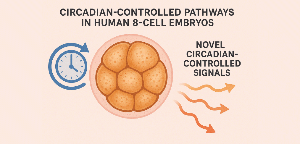
Our circadian rhythm studies continue to reveal unique characteristics of activated eggs. Research published in early 2024 by BRF scientists describes circadian-controlled pathways not previously known to function in activated human eggs. As a consequence, novel circadian-controlled signals released by the embryo have now been discovered.

These findings add to the growing evidence that biological rhythms play a more central role in early development than previously understood—and that timing itself may be a key variable in how we activate and sustain viable human cells in the lab.

This research also raises urgent questions for the field of cell culture: if circadian signals are active this early, we may need to take them into account to keep cells alive and healthy in the lab. It’s possible that many IVF clinics—and research labs—are using culture conditions that don’t reflect the biological timing cues embryos rely on.

One striking insight from the publication is the possibility that the 8-cell embryo may be secreting oxytocin, potentially to stimulate its own movement through the fallopian tube or to support early neural development. The study also confirms that the 8-cell embryo does not produce reproductive steroid hormones—an important finding that suggests hormonal signaling at this stage is likely driven by other pathways.

As seen in previous research on growth factors, the study also showed that the 8-cell embryo tends to produce more hormones than it has receptors for—suggesting that its signals may be directed outward, preparing for interaction with its environment rather than responding internally.

Taken together, these findings reveal a more dynamic picture of early human development—one in which communication, timing, and environmental readiness all play a role. Understanding these early signals could help scientists design better culture systems and may ultimately lead to improved outcomes in both research and clinical care.

Read the publication

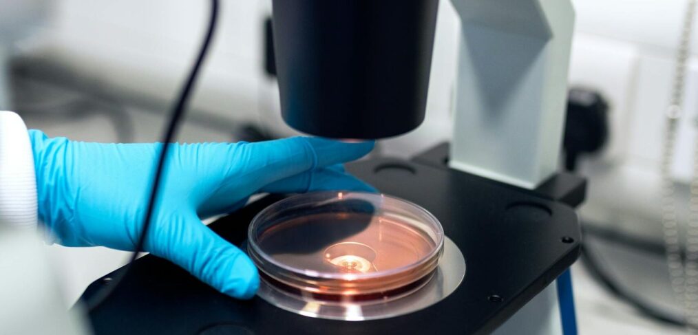
Scientist using a microscope to examine petri dish samples in a laboratory setting.

Zinc: A Possible Missing Ingredient for Incubation

As BRF scientists expand their access to research eggs, new findings from the broader scientific community are offering additional insights. One such development is the possible role of zinc in egg activation.

For many years, calcium ions have been known to play an important role in egg activation across many species and have been central to laboratory activation protocols. But recent research suggests that zinc may also be critical — and could offer a significant boost to incubation conditions that support development into continuously dividing parthenote stem cells.

BRF researchers are now working to incorporate this emerging science into future protocols.

Rescuing Discarded Eggs: A Breakthrough in Stem Cell Research

On the order of half of the human eggs collected during fertility procedures are not robust enough for reproductive use and are discarded by egg banks. BRF scientists developed a new human egg transport system that makes it possible to rescue and study these otherwise lost eggs for stem cell research.

As egg freezing has become more common, especially among younger donors, the number of eggs collected has grown, along with the number discarded for not meeting clinical standards. Until recently, it was difficult and costly to transport these unfertilized eggs to research laboratories due to their fragility. BRF new transport system marks a significant step forward on the path to universal parthenote stem cells for everybody.

The new Ambient Temperature Transport Medium (ATTM) is so novel and useful that patent protection is now in progress, following submission of a full research report on the findings. For the first time, eggs that could not previously survive transport to a lab can now be safely delivered for research.
This advance allows BRF to study an increased number of human eggs that were previously discarded — turning a lost resource into a vital one. These rescued eggs not only expand the Foundation’s ability to develop parthenote stem cells—an alternative to embryo-derived cells—but also provide valuable insight into human egg maturation more broadly.

Federal Funding Bans Won’t Stop Us

Independent Research is in Our DNA

The need for independent research has never been more evident. At the Bedford Research Foundation, the decades-old ban on federally funded stem cell research forced us to forge our own path—one that isn’t tied to federal funding. Now, as the important work of so many of our fellow researchers is hit hard due to massive federal cuts, our laboratory is able to carry foward the mission of basic science.

Organizations reliant on NIH funding often allocate 40-75% to overhead costs like administration and facilities, meaning that only a fraction of your donation directly supports research. For every dollar the NIH distributes, it costs taxpayers about $1.50-1.75, and then only about 60 to 25 cents goes toward research.

In contrast, at Bedford Research Foundation, 96 cents of every dollar you give goes straight toward basic research — research targeting breakthroughs in treating Alzheimer’s, Parkinson’s, neurodegenerative diseases, HIV, blood disorders, and cancers.

We’re proud to hold a four-star rating on Charity Navigator, and we’re home to a pioneering research team, whose visionary work continues to push the boundaries of what’s possible in medical science. Regardless of the political climate, our commitment to solving critical health challenges remains unwavering.

Your support today will make a transformative difference. Join us in funding the research that brings hope and cures to countless lives. You can change the life of everyBODY that you know.

JARG – Advancing Fertility Science: Explore the Latest Research in Assisted Reproduction and Genetics

Discover cutting-edge research and innovations in assisted reproduction technology and genetics. The Journal of Assisted Reproduction and Genetics offers the latest insights from reproductive biology to safe ART practices, helping shape the future of fertility treatments and genetic understanding.

Read the Journal of Assisted Reproduction and Genetics here.


BRF’s Latest CMV Data Suggests an Urgent Need for Better Understanding for Semen in IVF

Bedford Research Foundation scientists publish the latest CMV (cytomegalovirus) research data in the European Society of Medicine Journal.

Between April 2020 and March 2022, BRF collected 453 semen specimens from 156 HIV-infected men. The men were seeking specialized HIV PCR testing for semen provided by SPAR (the Special Program of Assisted Reproduction). Through SPAR, semen specimens with no detectable HIV viral load are cryopreserved and transferred to a partner clinic for use in fertility procedures. The goal is is to help couples achieve a pregnancy without transmitting the father’s infection to the mother, surrogate, or child.

In recent years, BRF has initiated CMV testing on all semen specimens going through SPAR. The testing was initiated because of the alarming reports from the CDC that CMV is now the leading infection cause of birth defects in the United States. It’s been known since at least 2007 that 1 in 750 babies born in the US has birth defects due to CMV. Additionally, although the CDC reports that between 50 and 80 percent of people in the United States have had a CMV infection by the time they are 40 years old, it is well-documented that  even as of 2024, very few couples are counseled about the disease during pregnancy.

Read More

Fertilized Eggs Versus Embryos, An Ongoing Failure of Terminology

“What is an Embryo?” Connecticut Law Review, 2004, with forward by Ruth Bader Ginsberg.

Nature does not regard fertilized human eggs as children. Nature’s stringent biological requirements of a fertilized egg leads to miscarriage far more often than successful pregnancy. Fewer than 10% of eggs fertilized in fertility clinics have the biological potential to develop into a child. Nature has evolved rigorous developmental milestones to avoid wasting precious maternal uterus time on a fertilized egg without the capacity for successful development to birth. It’s a numbers’ game. The nine months of human pregnancy means nine eggs will not have the opportunity to be released by the ovary for fertilization, so the fertilized egg occupying the maternal resources needs to prove it is worth the loss of nine other chances to continue the species. 

Nature does not regard fertilized human eggs as children.

Read More

What is an Embryo?

[Check out this month’s Science Highlights “Fertilized Eggs Versus Embryos, An Ongoing Failure of Terminology.]

In light of the recent news from Alabama, we are reposting “What Is An Embryo?”.

This is the first comprehensive look at the influence of accurate science terminology, published by the Connecticut Law Review along with rejoinders by Dr. Harold Shapiro, Prof John A. Robertson, Prof Lars Noah, and Father Kevin P. Quinn.

The law review addresses the controversy of all of the entities that are currently called “embryos” with regards to embryonic stem cell research legislation around the world.

Download the PDF: What Is an Embryo? 

“Ann Kiessling’s thoughtful and extremely useful review covers a good deal of history, language, and public policy intimately related to the controversial technologies that surround the use of embryos either in biomedical research or ART.”
– Harold T. Shapiro, Emeritus, Princeton University

Read More